


Size
| Size I.D. (mm) | Item No. |
| 6 | RET-S-6.0 |
| 6.5 | RET-S-6.5 |
| 7 | RET-S-7.0 |
| 7.5 | RET-S-7.5 |
| 8 | RET-S-8.0 |
| 8.5 | RET-S-8.5 |
| 9 | RET-S-9.0 |
Description
After the glottis was exposed under direct vision with the aid of laryngoscope, the catheter was inserted into the trachea through the mouth.
Tilt client's head back and lift jaw forward and upward with both hands to open mouth, or rotate mouth with right thumb pointing at lower teeth and pointing at upper teeth.
Hold the handle of the laryngoscope in the left hand, put the laryngoscope into the mouth through the right corner, push the tongue to the side, and then slowly advance to the uvula. Lift the lens vertically forward until the epiglottis is exposed. Raise the epiglottis to expose the glottis.
If intubation with curved lens is used, place the lens at the junction of epiglottis and tongue root (epiglottis valley), lift the lens forward and upward forcefully, so that the hyoid epiglottis ligament is tense, and the epiglottis is tilted close to the laryngoscope, that is, the glottis is exposed. If intubated with straight lenses, the epiglottis should be raised directly to expose the glottis.
Hold the middle and upper segments of the catheter with your right thumb, index finger and middle finger as if holding a pen, enter the mouth through the right corner of the mouth, and move the end of the catheter to the laryngoscope until the catheter approaches the laryngoscope. At the same time, monitor the direction of the catheter through the narrow gap between the lens and the tube wall, and accurately and lightly insert the tip of the catheter into the glottis. With the help of tube core intubation, when the tip of the tube enters the acoustic door, the tube core should be pulled out before the tube is inserted into the trachea. The depth of the catheter into the trachea for adults is 4-5cm, and the distance from the catheter tip to the incisor teeth is about 18-22cm.
After intubation is complete, ensure that the catheter is inserted into the trachea before fixation. Confirmation methods include:
When you press the chest, there's airflow through the catheter.
During artificial respiration, symmetrical fluctuation of bilateral thorax was observed and clear alveolar breath sounds could be heard.
If the transparent catheter is used, the tube wall is clear when inhaling, and obvious "white fog" changes can be seen when exhaling.
If the patient is breathing spontaneously, the breathing capsule may be seen to contract with breathing after the anesthesia machine.
If end-expiratory ETCO2 can be monitored, it will be easier to judge, and it can be confirmed if the ETCO2 graph is displayed [1].
The endotracheal tube is inserted into the trachea through the nasal cavity under blind vision.
Spontaneous breathing must be retained during intubation, and the direction of the catheter can be determined according to the strength of the exhaled air flow.
Use 1% tetracaine as intranasal surface anesthesia, and drop 3% ephedrine to make nasal mucosa vasoconstriction, in order to increase nasal volume, and can reduce bleeding.
Use an appropriately sized endotracheal tube and insert it into the nasal cavity with the right hand. During intubation, listen for the strength of exhaled air while moving forward, and adjust the patient's head position with the left hand to locate the strongest exhaled air.
The catheter is pushed forward quickly as the glottis opens. When the catheter entered the glottis, the propulsion resistance was reduced, and the exhaled airflow was obvious. Sometimes the patient had cough reflex. After the anesthesia machine was connected, the breathing capsule could be seen contracting with the patient's breathing, indicating that the catheter was inserted into the trachea.
If the exhaled air disappears after the catheter is advanced, it is inserted into the esophagus. The catheter should be retreated to the nasopharynx with the head slightly tilted so that the tip of the catheter is upturned and can be aligned with the glottis to facilitate insertion.
Products categories
-

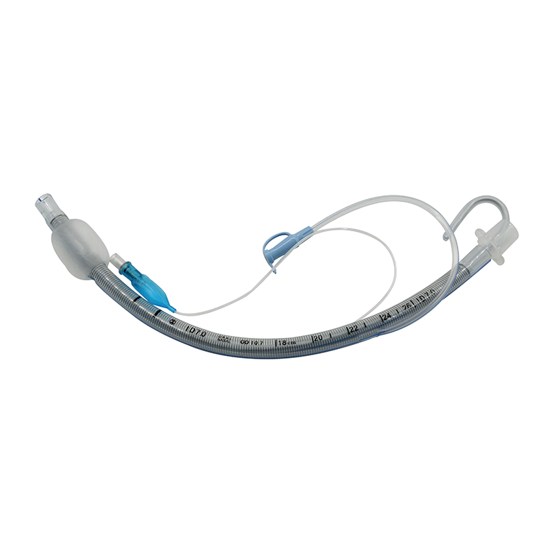
Standard Endotracheal Tube with suction lumen(S...
-

Oral Preformed Endotracheal Tube with cuff (OET-C)
-

Oral Preformed Endotracheal Tube without cuff (...
-

Nasal Preformed Endotracheal Tube with cuff (NE...
-

Standard Endotracheal Tube with suction and dos...
-

Endotracheal Tube with cuff(ETT-C)